Smith Lab

Learn About Electric Fish
A relatively simple neural circuit controls the EOD
One of the advantages of using electric fish to study the mechanisms of behavior is that the brain regions that control the EOD and its modulations are well-characterized and relatively simple compared to circuits that control sexually dimorphic behaviors in many other vertebrates. Furthermore, the activity of neurons in these brain regions is directly related to the EOD itself.
EOD frequency is controlled by the pacemaker nucleus (Pn) in the hindbrain. In the family of electric fish that we study (Apteronotidae), the pacemaker nucleus is located on the ventral surface of the brain, and is visible as a bump protruding from the bottom of the medulla.
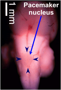 |
(Left) Ventral view of the brain of a brown ghost knifefish (A. leptorhynchus). The pacemaker nucleus, which controls the EOD, is visible as a protrusion on the ventral surface of the hindbrain (blue arrowheads). |
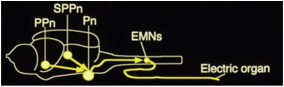
(Right, top) Sagittal diagram of the brain circuit that controls the EOD. The electric organ is formed from the axons of electromotor neurons (EMNs) whose cell bodies lie in the spinal cord. EOD frequency is controlled by the pacemaker nucleus. Chirping is controlled by the prepacemaker nucleus (PPn), and the jamming avoidance response is controlled by the subleminiscal prepacemaker nucleus (SPPn). (Right, bottom) Schematic illustration of cell types of the pacemaker nucleus and their connections to each other and the EMNs in the spinal cord. Based on Smith (1999). |
 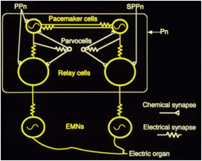 |
The pacemaker nucleus contains three types of neurons: pacemaker cells, relay cells, and parvocells. All of the pacemaker cells are electrically coupled to each other, which allows them to precisely and synchronously fire action potentials at high frequencies. The pacemaker cells are also electrically coupled to relay cells, which send their axons down the spinal cord. The relay cell axons form electrical synapses on electromotor neurons in the spinal cord. In apteronotid fish, the axons of electromotor neurons form the electric organ. The EOD in apteronotid fish is generated by the action potentials in the axons of the electromotor neurons.
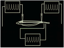
Action potentials of pacemaker and electromotor neurons correspond directly to the EOD.
The firing rates of pacemaker, relay, and electromotor neurons are directly related to EOD frequency. To generate each discharge of the electric organ, all of these neurons fire a single action potential. Thus, in a fish whose EOD frequency is 1000 Hz, all pacemaker, relay, and electromotor neurons produce 1000 action potentials each second. Any changes in the firing rates of these cells are translated directly into corresponding changes in EOD frequency. A major component of our research is understanding the cellular mechanisms that control the firing rates of neurons in the electromotor system; how these mechanisms vary across individuals, sexes, and species to produce diversity in EOD frequency; and how hormones alter those mechanisms to produce sex differences in EOD frequency.
Modulations of the EOD, including chirping and the JAR are controlled by two brain regions, the prepacemaker nucleus (PPn) in the thalamus and subleminiscal prepacemaker nucleus (sPPn) in the midbrain. Both the PPn and sPPn project to the pacemaker nucleus and form excitatory connections on pacemaker and/or relay cells. When neurons in the PPn fire, they release glutamate onto relay cells in the pacemaker nucleus. This causes the relay and pacemaker cells to fire faster for a fraction of a second, which in turn causes the transient increase EOD frequency that constitutes a chirp. Another component of our research examines how sex and species differences in the PPn and pacemaker nucleus are related to sex and species differences in chirping.