Smith Lab

The Electromotor System is a Model for Motor Pattern Generation
The electric organ discharge (EOD) and the regular firing of the neurons that control it are among the fastest and most precise biological oscillations.
In apteronotid electric fish, EOD frequency can approach 2000 Hz.The firing rates of pacemaker neurons and electromotor neurons correspond directly to EOD frequency, and these neurons thus produce action potentials synchronously at rates of up to 2000 times per second. One of the goals of our research is to understand the cellular mechanisms that underlie these rapid and precise motor patterns and to understand how variation in neuronal properties among individuals and between sexes and species is related to behavioral diversity.
Pacemaker and electromotor neurons continue to fire spontaneously in vitro.
Studying the cellular mechanisms of high-frequency motor rhythms is facilitated by the simplicity of the neural circuits that control the EOD and by the fact that these neurons maintain in vivo-like activity when recorded in vitro. (click here for background information on the electromotor circuit) The pacemaker nucleus and electromotor neurons can be removed from the animal and kept alive in artificial cerebrospinal fluid. These neurons maintain spontaneous firing rates that approximate the in vivo EOD frequency of the fish from which they were taken (see figure below). This allows us to use electrophysiological techniques to study these neurons under controlled, in vitro, conditions, but still be able to understand how variation in the activity of these neurons between individuals, sexes, or species is related to the in vivo electrical behavior of the fish.
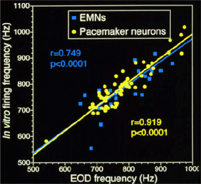 |
Scatter plot of EOD frequency in vivo and in vitro firing frequency of electromotor neurons (blue) and pacemaker neurons (yellow) in slice preparations. Note the strong correlation between in vivo and in vitro firing rates. Based on data from Meyer (1984); Schaefer and Zakon (1997); and Smith and Zakon (2000). |
We use two approaches to study mechanisms of high frequency motor pattern generation in pacemaker and electromotor neurons:
Experiments with Channel Blockers to Identify Ionic Currents that Control Spontaneous Firing
Because both pacemaker neurons and electromotor neurons continue to fire in vitro at frequencies very similar to those in vivo, we are able to record their spontaneous firing patterns to study how neuronal excitability influences behavior. We can then apply drugs that specifically block particular classes of ion channels to identify the ionic currents that generate the high-frequency firing patterns that control the EOD rhythm.
We have used this technique to study ionic currents that control high-frequency firing in both pacemaker neurons (Smith and Zakon 2000) and electromotor neurons (Smith 2006). These studies revealed that sodium and potassium currents are critical for generating and regulating the motor rhythm for the EOD: (1) fast, transient sodium currents rapidly generate the depolarizing phase of action potentials; (2) persistent sodium currents regulate firing rates and provide tonic depolarization to maintain high-frequency firing; and (3) potassium currents carried by Kv1-like channels contribute to the repolarization of the action potential and also regulate firing rates. We have also used immunohistochemistry to show that several different Kv1 potassium channels are expressed in the pacemaker nucleus and in the axons of electromotor neurons (Smith, et al. 2006).
Voltage-clamp Experiments to Characterize the Biophysical Properties of Ionic Currents in Pacemaker and Electromotor Neurons.
The studies described above revealed that the firing patterns of pacemaker and electromotor neurons are regulated primarily by voltage-gated sodium and potassium currents. A major goal of our research is to understand how the properties of these currents allow neurons in the electromotor system to fire rapidly, and ultimately, how these properties influence EOD frequency. We are investigating these questions by using the voltage-clamp technique.
We use a microscope with Nomarski optics and an infrared-sensitive video camera to visually guide fire-polished glass micropipettes onto the membranes of pacemaker or electromotor neurons. By applying suction to the micropipette, we form a gigaohm seal onto the membrane, and record the sodium and potassium currents flowing either through small numbers of ion channels (cell-attached or outside-out patch clamp) or across all of the ion channels in the cell (whole-cell recording). The voltage clamp amplifier allows us to control the voltage across the membrane and measure the ionic currents at different voltages. We can thus determine several important biophysical properties of these currents: (1) the voltages at which they activate; (2) the degree and voltage-dependence of inactivation; and (3) the speed of activation and inactivation. Measuring these parameters will allow us to generate a model of how currents interact to produce action potentials at rates of more than 1000 Hz. Ultimately, we can also compare the properties of ionic currents in pacemaker and electromotor neurons from fish with high-frequency EODs (e.g., male brown ghost knifefish) with those from fish with low-frequency EODs (e.g., female brown ghost knifefish) to understand how hormones act on ion channels in identified neurons to produce sex differences in behavior and how evolution has affected neuronal excitability to produce species diversity in behavior. These studies are allowing us to examine the mechanisms of sex and individual differences in behavior from the level of single cells to entire organisms, and to study how these mechanisms evolve to produce different behaviors across species.
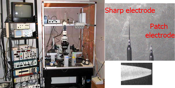 |
(Left) Electrophysiology station in the Smith lab is used for voltage- and current-clamp recordings of neurons in in vitro preparations of brain regions from the electromotor system. (Right) Recording electrodes for sharp intracellular recordings and patch clamp recordings. |